UMD Researchers Develop Tools to Sharpen 3D View of Large RNA Molecules
New technique breaks through a technology roadblock that limited RNA imaging for 50 years.
University of Maryland scientists have developed a method to determine the structures of large RNA molecules at high resolution. The method overcomes a challenge that has limited 3D analysis and imaging of RNA to only small molecules and pieces of RNA for the past 50 years.
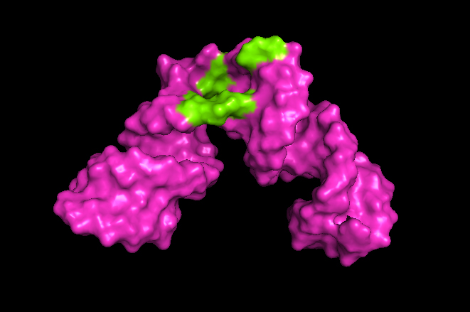
The new method, which expands the scope of nuclear magnetic resonance (NMR) spectroscopy, will enable researchers to understand the shape and structure of RNA molecules and learn how they interact with other molecules. The insights provided by this technology could lead to targeted RNA therapeutic treatments for disease. The research paper on this work was published in the journal Science Advances on October 7, 2020.
“The field of nuclear magnetic resonance spectroscopy has been stuck looking at things that are small, say 35 RNA building blocks or nucleotides. But most of the interesting things that are biologically and medically relevant are much bigger, 100 nucleotides or more,” said Kwaku Dayie, a professor of chemistry and biochemistry at UMD and senior author of the paper. “So, being able to break down the log jam and look at things that are big is very exciting. It will allow us to peek into these molecules and see what is going on in a way we haven’t been able to do before.”
To read more, visit https://cmns.umd.edu/news-events/features/4683